Blood (Plasma & Formed Elements)
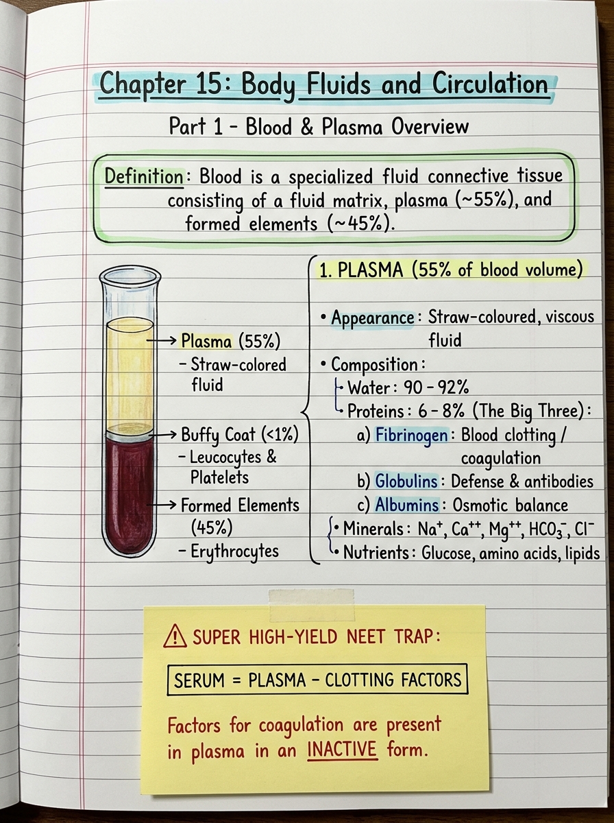
Definition: Blood is a specialized fluid connective tissue consisting of a fluid matrix, plasma (~55%), and formed elements (~45%).
1. Plasma (55% of Blood)
- Appearance & Nature: Straw-coloured, viscous fluid.
- Composition:
- Water: 90–92%
- Proteins (6–8%): The Big Three:
- Fibrinogen: Essential for blood clotting / coagulation.
- Globulins: Primarily involved in defense mechanisms (immunoglobulins/antibodies).
- Albumins: Maintain colloid osmotic balance (keeps water inside blood vessels).
- Minerals/Electrolytes: Na+, Ca2+, Mg2+, HCO3-, Cl-.
- Nutrients in transit: Glucose, amino acids, lipids.
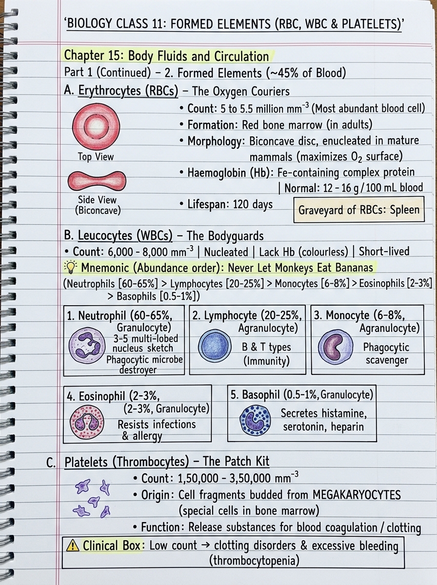
2. Formed Elements (45% of Blood)
A. Erythrocytes (RBCs) – The Oxygen Couriers
- Count: 5 to 5.5 million mm-3 of blood (most abundant).
- Site of Formation: Red bone marrow (in adults).
- Morphology: Biconcave disc, enucleated (no nucleus in mature mammalian RBCs — increases surface area for O2).
- Haemoglobin (Hb): Iron-containing complex protein. Normal value: 12–16 g per 100 mL of blood.
- Lifespan: 120 days.
- Graveyard: Destroyed in the spleen.
B. Leucocytes (WBCs) – The Bodyguards
- Count: 6,000–8,000 mm-3 of blood.
- Features: Colourless (no Hb), nucleated, relatively short-lived.
Never Let Monkeys Eat Bananas
Neutrophils > Lymphocytes > Monocytes > Eosinophils > Basophils
| Type | Classification | % of Total WBC | Key NCERT Function / Features |
|---|---|---|---|
| Neutrophil | Granulocyte | 60–65% (Most abundant) | Phagocytic cells (destroy invading microbes). Multi-lobed nucleus. |
| Lymphocyte | Agranulocyte | 20–25% | B & T forms; immune responses (antibodies & cell-mediated immunity). |
| Monocyte | Agranulocyte | 6–8% | Phagocytic; large kidney/bean-shaped nucleus. |
| Eosinophil | Granulocyte | 2–3% | Bilobed nucleus; resists infections & associated with allergic reactions. |
| Basophil | Granulocyte | 0.5–1% (Least abundant) | Secretes histamine, serotonin, heparin; involved in inflammatory reactions. |
C. Platelets (Thrombocytes) – The Patch Kit
- Origin: Cell fragments budded off from megakaryocytes (special giant cells in the bone marrow).
- Count: 1,50,000–3,50,000 mm-3.
- Function: Release coagulation factors for blood clotting.
- Clinical note: A drop in platelet count (thrombocytopenia) leads to clotting disorders & excessive blood loss.
Blood Groups & Coagulation Cascade
1. ABO Blood Grouping
Karl Landsteiner ne discover kiya tha. Blood group decide hota hai RBC ki surface par present Antigens se:
- Antigen: Chemical/protein jo RBC membrane ke bahar laga hota hai (induces immune response).
- Antibody: Blood plasma ke andar natural defence proteins (antigen ke against attack karne wale).
| Blood Group | Antigen on RBC surface | Antibody in Plasma | Can Donate To (Safe) | Can Receive Blood From (Donors) |
|---|---|---|---|---|
| A | A | anti-B | A, AB | A, O |
| B | B | anti-A | B, AB | B, O |
| AB | A and B | NIL (None) | AB only | AB, A, B, O (Universal Recipient) |
| O | NIL (None) | anti-A and anti-B | A, B, AB, O (Universal Donor) | O only |
- Donor ki RBC ka Antigen aur Recipient ke Plasma ki Antibody same nahi honi chahiye, warna clumping (agglutination) aur RBCs ka destruction ho jayega!
- Universal Donor (O-): Iske RBC surface par koi antigen nahi hai, isliye kisi bhi recipient ke antibodies ispe attack nahi kar paate.
- Universal Recipient (AB+): Iske plasma mein koi antibody nahi hai, isliye ye kisi ka bhi antigen accept kar leta hai.
2. Rh Grouping & Erythroblastosis Foetalis
- Rh Antigen: Pehle Rhesus monkey mein discover hua tha. Nearly 80% humans are Rh+ve, baaki 20% are Rh-ve.
- An Rh-ve person normally has no Rh antibodies, but agar Rh+ve blood se expose ho jaye, toh unke andar anti-Rh antibodies banne lagte hain.
🚨 Erythroblastosis Foetalis (The Classic NEET Question):
1. Pure 9 Mahine Tak (Placental Barrier):
Baby ka blood aur mother ka blood kabhi aapas mein direct mix nahi hote. Baby ki blood vessels Chorionic Villi ke andar band hoti hain. Beech mein ek semi-permeable membrane hoti hai (Placental Barrier). Sirf O₂, glucose, aur chote molecules diffuse hote hain. RBCs is barrier ko cross nahi kar sakti.
2. Parturition (Delivery) ke Time Achanak Kya Hota Hai? (The Breakage):
Baby ke bahar aane ke baad uterus zor se contract hota hai taaki placenta (jo uterine wall se chipka hua tha) ukhade aur alag ho. Placenta ke alag hone (shearing) par baby ki chorionic villi ki microscopic capillaries rupture (phat) jaati hain! Us tearing ke dauran, baby ki tooti hui capillaries se 1–2 mL foetal blood mother ki exposed veins mein leak ho jaata hai!
Analogy: Jaise deewar par tape chipki ho jisme do alag colored tubes hain. Jhatke se tape ukhadoge toh dono capillaries phatengi aur halka sa leak hoga.
3. Toh Phir 1st Baby Bach Kaise Gaya?
Delivery ke time jab baby bahar aaya, uske baad blood leak hua. Baby toh already safe delivery room mein hai! Mother ke immune system ko foreign Rh antigen ke against antibodies banane mein hafton (weeks) lagte hain (Primary Immune Response). Jab tak mother ki body mein Anti-Rh antibodies (IgG) taiyaar hote hain, tab tak pehla bachha already paida ho chuka hota hai! 1st baby ko kuch nahi hota!
4. Par 2nd Baby Ke Time Kaand Kyun Hota Hai?
Mother ki body ab "sensitized" ho chuki hai (Memory B-cells ban chuke hain). Jab 2 saal baad 2nd Rh+ve baby conceive hota hai, toh wo abhi pet ke andar hi hota hai. Maa ke blood mein pehle se bani hui Anti-Rh antibodies (IgG) placenta ko cross karke pet ke andar hi 2nd baby ke blood mein ghus kar uski RBCs ko phodna (hemolysis) shuru kar deti hain! Isse 2nd baby ko severe anaemia aur jaundice ho jaata hai.
Prevention (Clinical Fix): Mother ko Anti-Rh antibodies (RhoGAM injection) deliver kiye jaate hain immediately after the delivery of the 1st child (within 72 hours). Ye leak hue foetal cells ko neutralize kar deta hai isse pehle ki mother ka immune system memory banaye!
3. Coagulation of Blood (Clotting Cascade)
Cut lagne par shareer ko chhed band karne ke liye ek jaali (mesh/net) chahiye jisme cells phas jayein. Agar wo jaali pehle se khoon mein ghum rahi hoti, toh vessels block ho jaati aur insaan mar jaata! Isliye jaali banane wale saare karigar khoon mein SO RAHE HOTE HAIN (Inactive State). Cut lagne par chain reaction (Cascade) se jagte hain!
- Potassium (K⁺ ion): ❌ KOI ROLE NAHI HAI! Ye nerve impulse aur heartbeat ke liye hota hai. Clotting cascade se iska koi lena-dena nahi hai!
- Calcium (Ca²⁺ ion): EXTREMELY IMPORTANT! Yehi wo akela main mineral ion hai jo clotting reaction (Cascade) karwata hai (Factor IV).
- Vitamin K (Letter 'K'): VERY IMPORTANT! Ye koi mineral nahi hai, ye Vitamin hai! Liver ko Prothrombin banane ke liye Vitamin K ki zaroorat hoti hai.
Lymph (Tissue Fluid) & Circulatory Pathways
1. Lymph (Tissue Fluid / Interstitial Fluid)
Jab arterial blood tissue capillaries ke patle chhedon se guzarta hai:
- High pressure ki wajah se pani + chhote water-soluble substances bahar nikal kar cells ke beech ke spaces (interstitial spaces) mein aa jaate hain.
- Badi cheezein jaise bade proteins aur RBCs blood vessel ke andar hi reh jaati hain.
- Is nikalne wale fluid ko kehte hain: Interstitial Fluid ya Tissue Fluid (Minerals exactly same as Plasma!).
- Cells aur blood ke beech saara gas exchange (O₂, CO₂) aur nutrient exchange isi tissue fluid ke through hota hai ("Middleman of the body").
- Is fluid ko lymphatic system collect karke major veins mein drain kar deta hai. Is fluid ko LYMPH kehte hain.
1. Colour: Colourless fluid (kyunki RBCs nahi hoti!).
2. Specialised Lymphocytes: Immune responses ke liye responsible.
3. Fats Absorption (⚠️ Super Favorite Question): Fats intestinal villi ke andar present lymphatic capillaries jinko Lacteals kehte hain, unke through absorb hote hain!
2. Circulatory Pathways (Open vs Closed)
| Feature | Open Circulatory System | Closed Circulatory System |
|---|---|---|
| Pipes / Network | Blood pumped by heart passes through large vessels into open spaces or body cavities called sinuses. | Blood pumped by heart is always circulated through a closed network of blood vessels. |
| Cell Bathing | Organs/Tissues direct blood mein bathed rehte hain. | Cells direct contact mein nahi aate (tissue fluid beech mein hota hai). |
| Regulation | Flow cannot be precisely regulated (low pressure). | Flow of fluid can be more precisely regulated (considered more advantageous). |
| Examples (NEET) | Arthropods & Molluscs. | Annelids (Earthworm!) & Chordates. |
3. Evolution of Vertebrate Heart (Chamber Evolution)
| Class / Animal | Chambers | Atria & Ventricles | Type of Circulation | Blood Flow Pattern |
|---|---|---|---|---|
| Fishes | 2 | 1 Atrium + 1 Ventricle | Single Circulation | Heart pumps only deoxygenated blood to gills → oxygenated to body → deoxygenated returned to heart (Venous Heart). |
| Amphibians & Reptiles (except crocodiles) | 3 | 2 Atria + 1 Ventricle | Incomplete Double Circulation | Left atrium receives oxygenated blood, right gets deoxygenated blood. Both get mixed in the single ventricle which pumps out mixed blood. |
| Crocodiles 🐊 (Exception!), Birds 🦅 & Mammals 🫀 | 4 | 2 Atria + 2 Ventricles | Complete Double Circulation | Oxygenated and deoxygenated blood received by left and right sides without any mixing up. Two separate circulatory pathways! |
Human Heart Anatomy & Conducting System
1. Human Heart Anatomy
- Origin: Mesodermally derived organ.
- Location: Thoracic cavity mein, between two lungs, slightly tilted to the left. Size of a clenched fist.
- Covering: Protected by double-walled membranous bag, pericardium, enclosing pericardial fluid (reduces friction).
- 4 Chambers: Two small upper atria and two larger lower ventricles. (Ventricle walls are much thicker than atria; Left ventricle wall is the thickest!).
- The 3 Septa:
- Inter-atrial septum: Thin, muscular wall separating right and left atria.
- Inter-ventricular septum: Thick-walled partition separating left and right ventricles.
- Atrio-ventricular (AV) septum: Thick fibrous tissue separating atrium and ventricle of the same side.
* TRY = Tricuspid valve (3 muscular flaps/cusps) → Right Atrium aur Right Ventricle ke beech!
* BUY = Bicuspid valve / Mitral valve (2 muscular flaps) → Left Atrium aur Left Ventricle ke beech!
* Semilunar Valves: Right ventricle ke opening into Pulmonary artery aur Left ventricle ke opening into Aorta par present hote hain.
* Chordae Tendinae: Fibrous cords that anchor the flaps of AV valves to the papillary muscles in the ventricular walls, preventing valves from inverting/prolapsing into atria during ventricular systole!
2. Specialised Nodal Tissue (Conducting System)
Heart is Myogenic (Autoexcitable — ability to generate action potentials without any external stimuli).
| Nodal Tissue | NCERT Exact Location | Key Function / Feature |
|---|---|---|
| SAN (Sino-Atrial Node) | Right upper corner of Right Atrium | "The Pacemaker". Generates maximum action potentials (70–75 min-1, average 72). Responsible for initiating & maintaining rhythmic contractile activity. |
| AVN (Atrio-Ventricular Node) | Lower left corner of Right Atrium (close to AV septum) | Relays the action potential from atria to ventricles. |
| Bundle of His (AV Bundle) | Continues from AVN → crosses AV septum → top of interventricular septum | Divides into right and left bundles. |
| Purkinje Fibres | Spread throughout ventricular musculature | Delivers impulse rapidly to ventricular walls causing simultaneous contraction. |
Cardiac Cycle, Heart Sounds, Volumes & ECG
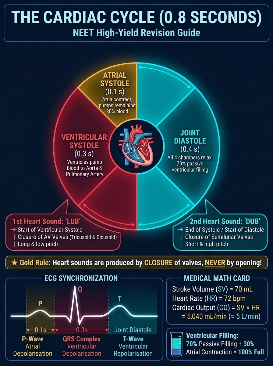
1. Cardiac Cycle (Total Duration = 0.8 Seconds)
Duration of 1 Cardiac Cycle = 60 seconds / 72 beats ≈ 0.8 seconds.
Ventricle ko 100% bharna hai:
1. Joint Diastole (0.4 s): Glass aur jug aaram se rakhe hain, pipe khula hai. Paani apne aap (gravity/passive flow se) behta rehta hai. Bina mehnat ke ventricle 70% bhar chuka hai!
2. Atrial Systole (0.1 s): Ab glass ko pura bharne ke liye 30% bacha hai. Atria zor se nichodta (contract hota) hai aur bacha hua 30% blood ventricle mein push kar deta hai!
70% (Passive flow) + 30% (Atrial push) = 100% Full Ventricle! Kuch bhi gayab nahi hua!
Dono Chambers ka Complete Time Breakdown (0.8 Seconds Total):
| Chamber | Systole (Contraction) | Diastole (Relaxation) | Total Time |
|---|---|---|---|
| Atria (Upper Chambers) | 0.1 s | 0.7 s | 0.8 s |
| Ventricles (Lower Chambers) | 0.3 s | 0.5 s | 0.8 s |
| Joint Diastole (Both relaxed together) | 0.4 s | 0.8 s | |
2. Heart Sounds (LUB vs DUB)
Stethoscope se aane wali aawaz valves ke BAND HONE se aati hai (Khulne ki koi aawaz nahi hoti!).
| Sound | Name | Kaunsa Valve Band Hua? | Kab Aati Hai? |
|---|---|---|---|
| 1st Sound | LUB | Tricuspid & Bicuspid (AV Valves) ke band hone par | Beginning of Ventricular Systole |
| 2nd Sound | DUB | Semilunar Valves ke band hone par | Beginning of Ventricular Diastole (End of systole) |
3. Cardiac Volumes & The "Each Ventricle" Trap
- Stroke Volume (SV): Volume of blood pumped out by each ventricle per cardiac cycle ≈ 70 mL.
- Cardiac Output (CO): Volume of blood pumped out by each ventricle per minute:
Cardiac Output = Stroke Volume × Heart Rate = 70 mL × 72 bpm ≈ 5000 mL or 5 Litres/min
5000 mL/min is pumped out by EACH VENTRICLE individually!
* Right Ventricle: Pumps 5 Litres/min into the Lungs.
* Left Ventricle: Pumps 5 Litres/min into the Body Tissues.
Dono 2.5–2.5 litre pump nahi karte! Double circulation series loop mein chalti hai, isliye dono ventricles 5 Litre each pump karte hain!
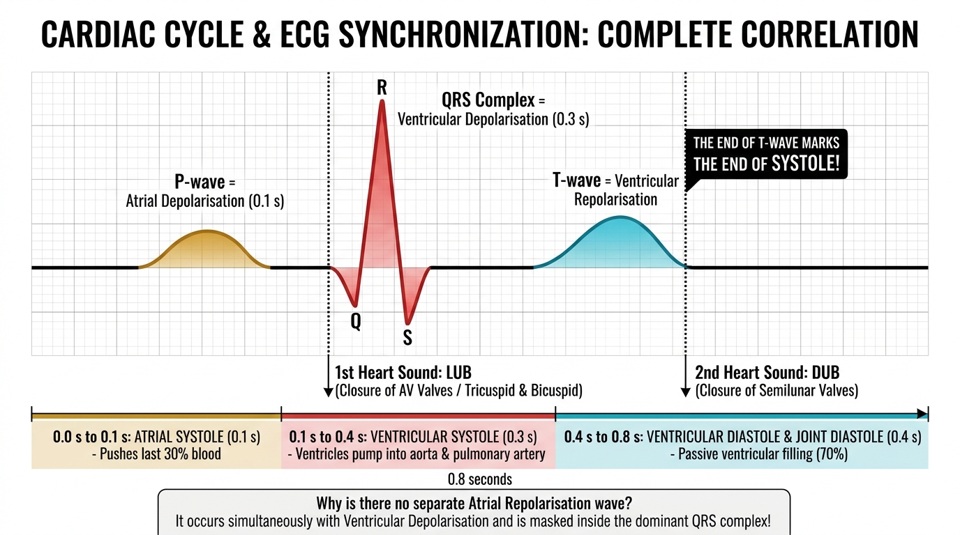
4. Electrocardiogram (ECG) & Synchronization
- Electro-cardiograph: Yeh MACHINE ka naam hai!
- Electro-cardiogram (ECG): Yeh GRAPHICAL RECORDING ka naam hai!
- Standard Leads: 3 electrical leads (one to each wrist + one to the LEFT ANKLE).
* P-wave: Atrial Depolarisation (Electrical excitation) → leads to Atrial Systole.
* QRS Complex: Ventricular Depolarisation → initiates ventricular contraction (contraction starts shortly after Q, AV valves close → LUB sound).
Where is Atrial Repolarisation? Atria repolarisation occurs at the same time as ventricular depolarisation, and is completely buried/masked inside the massive QRS complex!
* T-wave: Ventricular Repolarisation (Ventricles return from excited to normal state).
* "The end of the T-wave marks the end of systole!": Jaise hi T-wave ki tail khatam hoti hai, ventricles ka blood phekna officially finish ho jaata hai aur muscle relax hone lagti hai → Semilunar valves slam shut → DUB sound!
Double Circulation, Regulation & Circulatory Disorders
1. Structure of Blood Vessels (Arteries vs Veins)
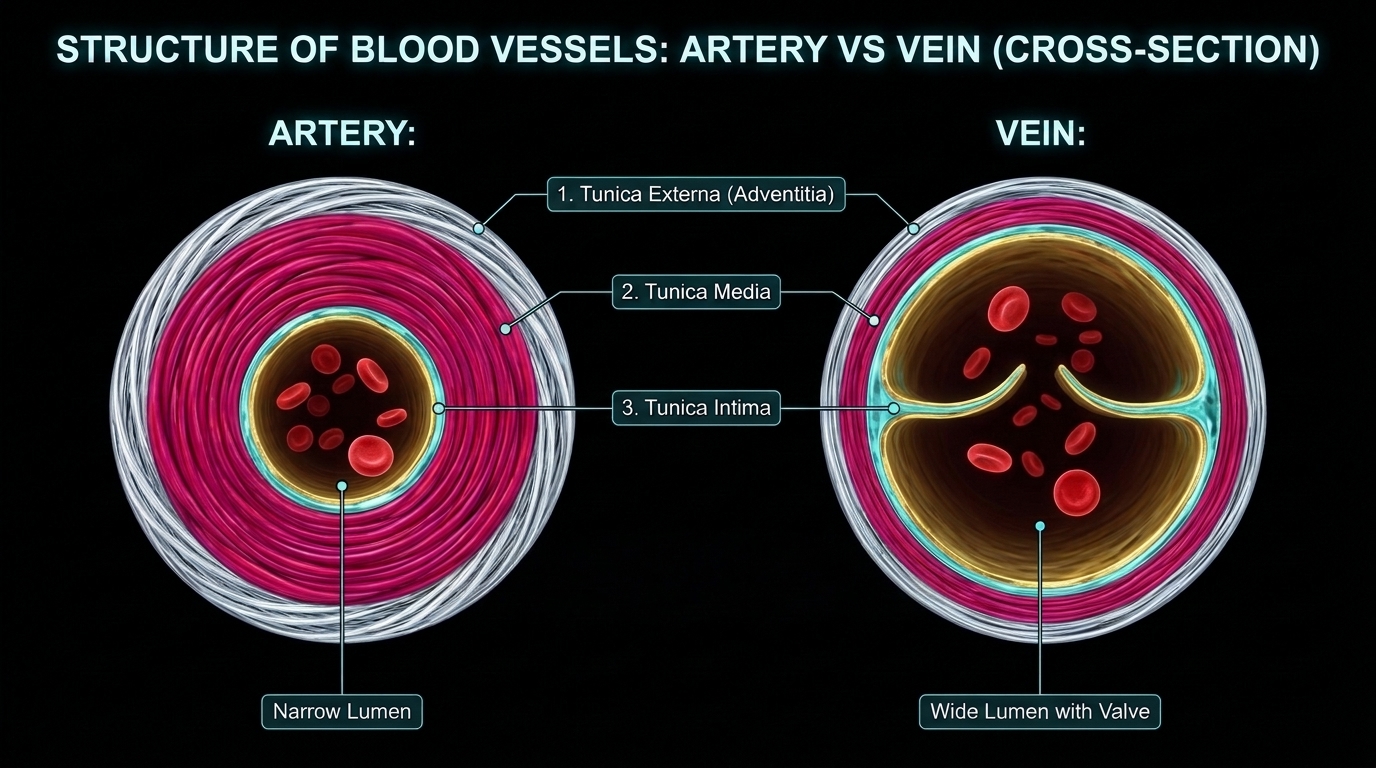
Artery ho ya Vein, dono ki deewar mein 3 layers (Tunics) hoti hain (Outer to Inner):
Artery: Moti deewar (thick tunica media) → Narrow Lumen, high pressure.
Vein: Patli deewar (thin tunica media) → Wide Lumen, low pressure + has Valves to prevent backflow.
2. Double Circulation (2 Separate Loops)
- Pulmonary Circulation: Right Ventricle (Deox) → Pulmonary Artery → Lungs → Pulmonary Veins → Left Atrium (Ox).
- Systemic Circulation: Left Ventricle (Ox) → Aorta → Arteries/Capillaries/Tissues → Veins/Vena Cava → Right Atrium (Deox).
3. Special Circulatory Systems
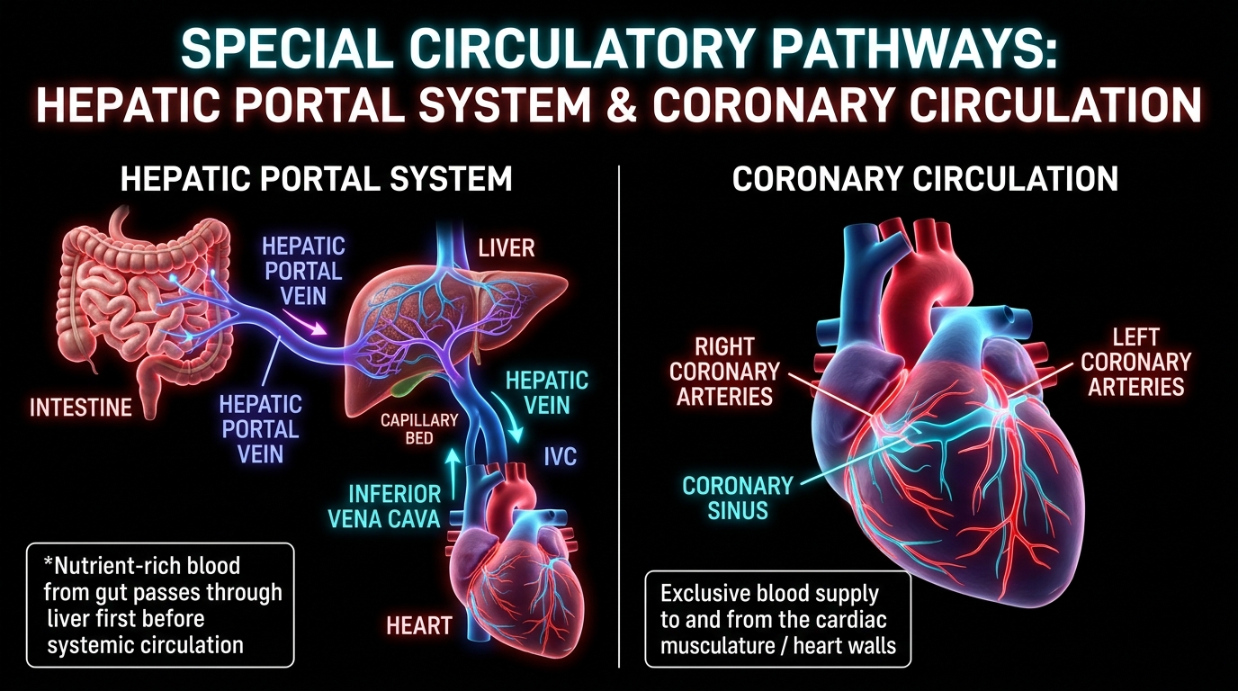
- Hepatic Portal System: Unique vascular connection between digestive tract and liver. Hepatic Portal Vein carries blood from intestine to liver before it is delivered to systemic circulation (metabolizes nutrients & detoxifies). Drains via Hepatic Vein into Inferior Vena Cava.
- Coronary System: Special system of blood vessels exclusively for circulation to and from cardiac musculature (heart walls). Aorta → Right & Left Coronary Arteries → Heart Muscles → Cardiac Veins → Coronary Sinus → Right Atrium.
4. Regulation of Cardiac Activity
- Myogenic: Auto-regulated intrinsically by nodal tissue (SAN).
- Neural Regulation: Medulla Oblongata moderates cardiac function through Autonomic Nervous System (ANS):
- Sympathetic Nerves: Increase heart rate, strength of ventricular contraction, and cardiac output.
- Parasympathetic Nerves: Decrease heart rate, decrease speed of conduction of action potential, and decrease cardiac output.
- Hormonal Regulation: Adrenal Medullary Hormones (Adrenaline / Noradrenaline) increase cardiac output.
5. Disorders of Circulatory System (The Big Four)
- Hypertension (High BP): Normal = 120/80 mm Hg (120 systolic, 80 diastolic). If repeated checks are ≥ 140/90 mm Hg → Hypertension. Damages vital organs like brain and kidney.
- Coronary Artery Disease (CAD) / Atherosclerosis: Deposits of calcium, fat, cholesterol, and fibrous tissues in vessels supplying heart muscle, making the arterial lumen narrower.
- Angina (Angina Pectoris): Symptom of acute chest pain appearing when not enough oxygen is reaching the heart muscle. More common in middle-aged and elderly.
🚨 The Big Three Differences (Heart Failure vs Cardiac Arrest vs Heart Attack):
| Condition | Exact NCERT Definition / Meaning |
|---|---|
| Heart Failure | State of heart when it is not pumping blood effectively enough to meet the needs of the body. Also called Congestive Heart Failure because congestion of the lungs is one of the main symptoms. |
| Cardiac Arrest | When the heart stops beating completely! (Flatline!). |
| Heart Attack (Myocardial Infarction) | When the heart muscle is suddenly damaged by an inadequate blood supply (blocked coronary artery). |
NCERT Summary & Page-by-Page Audit Gems
- "Beat Volume": Summary mein NCERT Stroke Volume ko "stroke or beat volume" kehti hai!
- Closed Circulation Generalization: Summary states: "All vertebrates and a few invertebrates have a closed circulatory system."
- Sponges & Coelenterates (Page 1): Simple organisms that lack blood; they circulate water from their surroundings through their body cavities for substance exchange.
- Chordae Tendinae (Page 6 Diagram): Anchor AV valves to papillary muscles of ventricles, preventing them from inverting during systole.
- Parasympathetic Mechanism: Specifically slows down the speed of conduction of action potential through the nodal system.
- Lymph Comparison: "Almost similar to blood except for the protein content and the formed elements."